We'd like to congratulation to our colleague Jarmila Stanková for defending her thesis Biologically active compounds’ molecular target localization and identification by microscopic methods. The thesis studied conjugates of biologically active substances, especially derivatives of betulinic acid with BODIPY. The study also presents a 3HQ-aminoBODIPY system with time-dependent drug release and ternary conjugates with real-time fluorescence change. An advanced analysis of the image dataset was performed, using artificial intelligence to classify topoisomerase inhibitors with high accuracy. This work suggests the promising use of conjugates in medicine and confirms the effectiveness of image data analysis using AI.
Mentor
Petr Džubák
Abstract
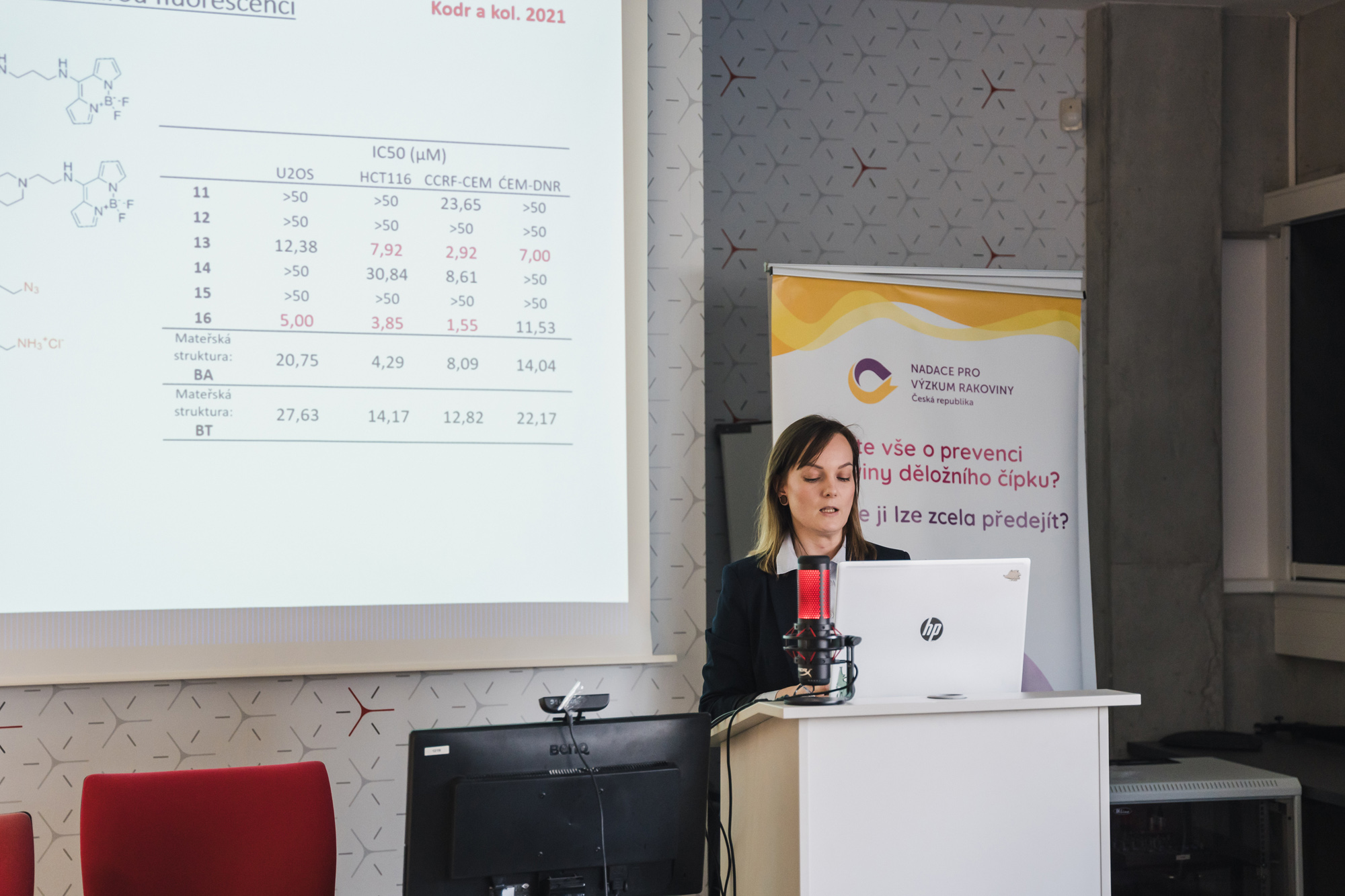
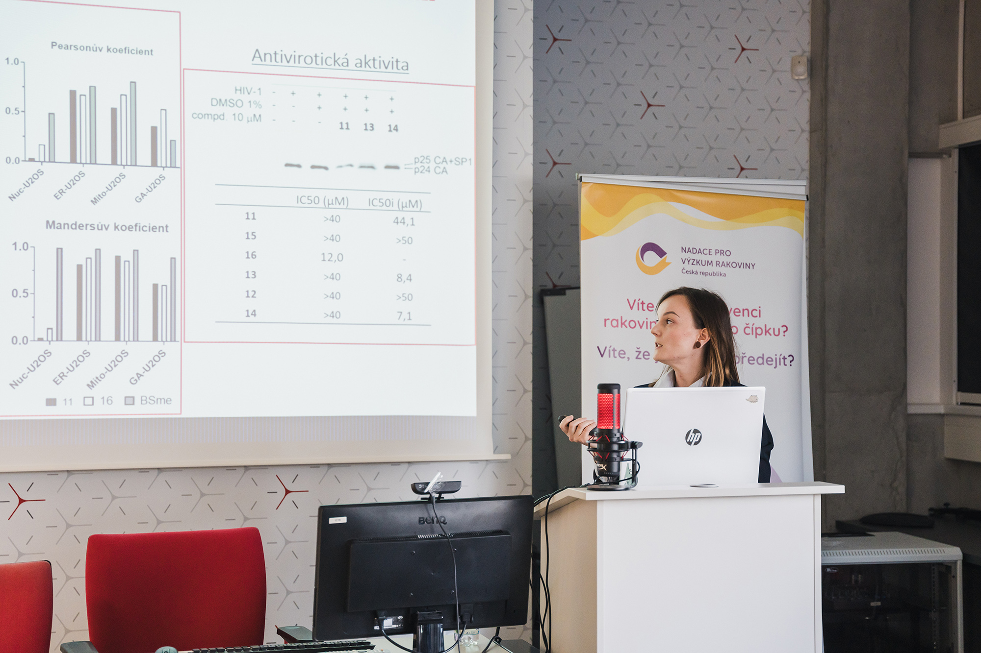
In this work, two distinct groups of conjugates of biologically active compounds with different variants of BODIPY dye were studied by fluorescent microscopy. Additionally, the research focused on a proof-of-concept study for the potential of a phenotypic screening method. Betulinic acid (BA) derivatives were tested on the U2OS cell line, where the first set of compounds included nine BA derivatives labeled with BODIPY-FL (conjugates 1-9), with a control conjugated containing only the linker and the BODIPY-FL label (conjugate 10). After a short incubation, these conjugates were localized in live cells. Conjugate 2 showed the highest cytotoxicity and a different staining pattern. The combination of the linker length and the triterpenoid moiety were crucial for the localization and the cytotoxicity of the conjugates. Visualization of the conjugates in fixed cells and cell lysates suggested possible covalent binding of the derivatives to cellular targets. The second set of compounds consisted of six BODIPY-labeled BA and bevirimat derivatives with blue fluorescence. A short incubation revealed the localization of conjugates 11 and 16 in living cells. Colocalization experiments showed interaction predominantly with the endoplasmic reticulum and mitochondria, emphasizing the importance of the added amino group.
The second part of the work focused on the 3HQ-aminoBODIPY system with OFF-ON effect to monitor the release of the active drug in cells. A disulfide linker was designed to regulate drug release in the presence of glutathione. Experiments with live cells and fluorescence microscopy confirmed the time-dependent effect of drug release from conjugates 17-20. The concept of three-part conjugates 20 and 21 was validated using the c(RGDfK) motif and FRET, where both conjugates were applied to HeLa cells, and the change in fluorescence was observed in real-time. This study highlights the potential of creating theranostics tailored for personalized medicine. In the final section of the work, an advanced analysis of the image dataset acquired by digital phase contrast (DPC) and fluorescence microscopy was performed. The data was analyzed by the commercial program Columbus and artificial intelligence (AI) using a convolutional neural network, allowing classification of topoisomerase inhibitors on the U2OS cell line with an accuracy up to 99%. This confirms the utility of DPC images for AI classification and their application in cell target identification and drug development.
Full information is available on theses.cz.
Photo: Denisa Pavelková